SKIN CANCER AWARENESS MONTH 2021
Skin cancer awareness month takes place every May with the aim of raising awareness of the dangers of unprotected sun exposure and educating on the ways skin cancer can be prevented. During skin cancer awareness month, the British Association of Dermatologists (BAD) also runs a sun awareness campaign encouraging people to self examine their body for skin cancer and to teach people about the dangers of sunburn and excessive tanning. This year, the national sun awareness week will run from Monday 3rd to Sunday 9th May.
SKIN CANCER
There are three main types of skin cancer, basal cell carcinoma (BCC), squamous cell carcinoma (SCC) and melanoma. The first two are both known as non-melanoma skin cancer and are the most common varieties.
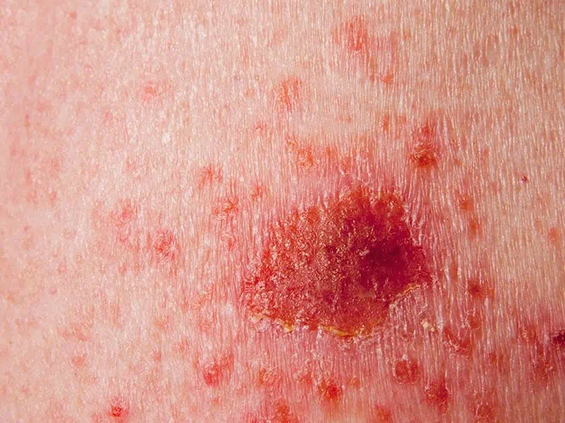
BASAL CELL CARCINOMA
Basal cell carcinoma forms in the cells which line the epidermis layer of the skin and accounts for around 75% to 80% of all skin cancers. Basal cell carcinoma is very unlikely to spread to other areas of the body and can be treated effectively. The earlier it is diagnosed and treated the better, as if left untreated, skin cancer can be fatal. The incidence of all types of skin cancer, including basal cell carcinoma, has increased dramatically over the last few decades. As with all skin cancers, exposure to ultraviolet (UV) light in the forms of sunlight or artificial tanning is the major cause of most BCCs.
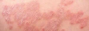
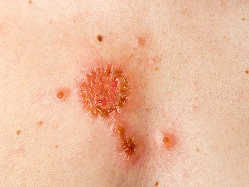
SQUAMOUS CELL CARCINOMA
Squamous cell carcinoma is less common and accounts for approximately 20% of all non-melanoma skin cancer. Most Squamous cell carcinomas can be cured, however a small amount can spread to other areas of the body, particularly the lymph nodes. Like lots of skin cancers, SCCs can vary in appearance. Most commonly, SCCs appear as a firm, raised lump which has a red, inflamed base and a rough, crusted surface. They tend to grow over time and can develop into an ulcer. The lump often feels sore or tender and can bleed. They can appear anywhere on the body, but most commonly found on areas of the body which are more frequently exposed to sunlight, including the face, head, neck and ears.
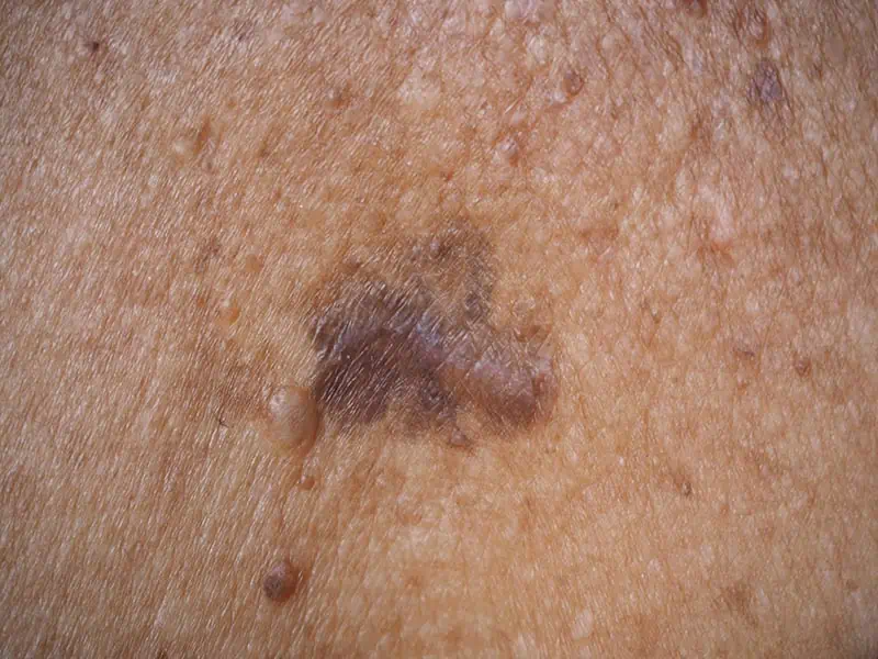
MELANOMA
Malignant Melanoma is less common, accounting for approximately 3% of all skin cancer cases in the UK, which equates to around 16,000 new cases a year. Although less common, malignant melanomas are very serious as they can grow quickly and spread to other organs in the body so it is vital they are diagnosed and treated early. Melanoma is a skin cancer that usually starts in the skin and develops when skin cells begin to develop abnormally.
It can start in a mole or even in normal looking skin and develops when melanocyte cells, the cells that produce brown pigment and moles, grow and divide at a quicker rate than usual.
As with all skin cancers, exposure to ultraviolet light (UV light) is the common cause. You also have an increased risk to developing malignant melanoma if you have lots of moles or freckles. If you have a pale skin type which is prone to burning or have a family history of the disease you are also more at risk.
The appearance of a new mole, or any changes to an existing mole could be an indication of a melanoma. This is why it is so important to regularly check and monitor your moles for any changes. In most cases, melanomas are more than one colour and are irregular in shape, they can sometimes be itchy and bleed.
HOW MUCH DO WE REALLY KNOW ABOUT MOLE CHECKING?
To support skin cancer awareness month, The Dermatology Partnership, a national network of start of the art dermatology facilities, of which Stratum Dermatology clinics are part of, has conducted research to understand the mole checking behaviours of the British public and to gauge how well we really know our moles and the warning signs of melanoma skin cancer.
It is recommended that moles are checked at home once a month, especially if you have lots of moles or freckles, have fair hair or skin, regularly expose your skin to sun or have a family history of skin cancer. When surveyed, those who have moles were found to not be checking as regularly as recommended, with 51% not checking their moles monthly. Of these, 24% of people either never check their moles for signs of skin cancer or go more than a year without checking their moles at home. Encouragingly, over a quarter of those surveyed (27%) check their moles daily or weekly, exceeding the recommended guidance.
When checking your own moles at home, there are particular things to be looking out for which could indicate malignant melanoma. These include changes to the size, shape and colour of moles. 45% of people surveyed said they were not confident that they knew the signs of skin cancer or what to be looking out for during a self mole check and 42% of people were only somewhat confident. Only 13% of those surveyed are very confident in knowing what to look for when checking their moles for cancer. This is a worrying statistic and shows that more work needs to be done in educating the public on how to check for moles.
HOW TO SELF CHECK YOUR MOLES
When checking your moles, it is recommended to use both a full length and a handheld mirror so you are able to check your body all over. Stand in a well-lit room and ask a family member or your partner to help you check the hard to reach areas such as your back.
Don’t forget to check less obvious places such as your scalp, the soles of your feet and in between your fingers and toes. It is a good idea to take photos of your moles so you can keep track of any changes.
Moles can look very different to each other which can make it hard to identify moles which may be worrying, but the key thing you are looking for is if your mole is changing. When checking your body for moles, you are looking for any changes to the size, colour of shape. You are also looking for itching, bleeding or crusting of moles which are signs you need to book an appointment to get your moles checked by a consultant dermatologist.
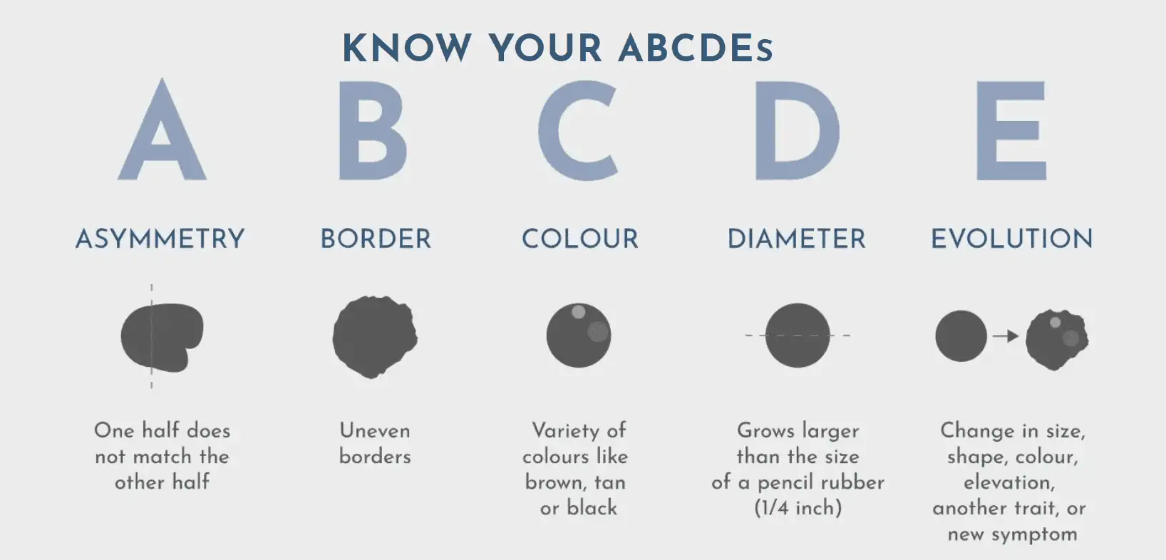
To help with this, an easy to remember acronym is often used for home mole checks to help you identify any worrying mole changes.
A: Asymmetry – moles should be symmetrical and a common sign of melanoma is an asymmetrical mole. If you were to draw a line through the middle of your mole or lesion and the two halves don’t match, then you should get it checked by a dermatologist. A common benign mole is often round or oval and symmetrical.
B: Border – if your mole has uneven borders then it could be malignant. Borders to look out for are uneven and may have scalloped or notched edges. Benign moles usually have smoother, even borders where the mole ends. If your mole shape changes to have an uneven border this could be a sign of melanoma.
C: Colour – if a single mole contains multiple colours within it when it could be a cause for concern. Benign moles are usually a single shade of brown, whereas melanoma may have different shades of browns, tan or blacks within it. Red, white and blue colours have also been identified in malignant moles and mole colour changes should be looked out for.
D: Diameter – malignant moles are likely to be larger than benign moles. A warning sign is when a mole or lesion is about the size of an eraser on the top of a pencil, about 6mm or 1.4 inch in diameter. This can be measured at home during your mole check routine and mole size changes should be noted. It is ideal to detect melanoma when it is small and in its early stages so it is important to not let your mole grow too big before you seek the services of a mole check clinic for a professional check.
E: Evolution – if your mole is changing, or evolving in any way over time then it may be cancerous. Any change in its size, shape, colour or evolution may be a warning sign, as well as any new symptoms seen inside the mole such as bleeding, itching or crusting of the skin.
It is a good idea to take regular photos of your moles at home so any of these evolutions or changes to your moles can be quickly identified and you can get a professional dermatologist mole check.
Worryingly, almost a quarter of those surveyed (23%), revealed that they have ignored a mole that they were worried about and not seeked professional advice. There are around 16,202 cases of melanoma skin cancer diagnosed every year in the UK, 86% of these cases are preventable and there is an 87% survival rate if treated quickly. This highlights the importance of regularly checking your moles and seeking professional diagnosis and treatment if required.
WHEN SHOULD YOU VISIT A DERMATOLOGIST FOR PROFESSIONAL MOLE CHECKING?
If you notice any suspicious changes in a mole, such as the ones outlined above, you should see an expert dermatologist for a professional mole check as soon as possible. Your dermatologist will make a detailed assessment of your worrying mole and either identify it as benign or recommend it to be removed.
Research has shown that professional mole checks by dermatologists catch melanoma skin cancer when it is thinner in size and earlier in its development, meaning there is a much higher survival rate. Melanoma caught early has about a 99% survival rate of five years, whereas if you catch it later, this rate can drop to around 60%.
If the mole needs to be removed, it will be tested in the lab to confirm whether it was cancerous and that all of the cancer cells have been successfully taken away. Sometimes, additional treatment will be required to eliminate any remaining cancer cells. In a small number of cases, the cancer will have spread to other parts of the body before it was detected. At this point, further treatment options will be discussed with you.
Noticing that one of your moles is changing can be worrying, but it is important to remember that most moles won’t cause any problems and most people won’t be affected by skin cancer.
If you have checked your moles and still aren’t sure if they need checking by a consultant dermatologist, take our interactive quiz to see if your moles are likely to be safe or need to be checked by an expert. If you have any doubts or concerns about your moles, book an appointment to get them checked. If caught and treated early, there is a 100% chance of survival.
HOW DOES A DERMATOLOGIST CHECK MOLES?
A professional mole check at a mole check clinic can be hugely reassuring as they can help to diagnose melanoma skin cancer early, which is vital for quick treatment and better long term outcomes. A dermatologist mole check is also an opportunity to get professional advice on how to check your moles at home and how to protect your skin when out in the sun.
Dermatologists have specialist training in dermatology, dermoscopy and pattern recognition, allowing them to differentiate benign (harmless) moles from dysplastic (abnormal) moles and melanoma (skin cancer). This allows early removal of dangerous moles and prevents unnecessary removal of benign moles.
There are a couple of different mole check services offered in a mole check clinic which are far more detailed and advanced than can be achieved with an at home mole check:
SINGLE MOLE CHECK
If you have a single mole which you are concerned about it can be checked in isolation. The mole will be scanned with a dermatoscope by a specialist nurse which will then be sent to a consultant dermatologists to be reviewed. A digital report on the mole will be produced and you will be contacted if further investigation is needed.
FULL BODY MOLE CHECK
During your dermatologist full body mole check, you will be seen by an expert consultant dermatologist. They will examine your skin using a dermatoscope, focusing on every mole and lesion. A dermatoscope uses a high-quality lens for 10 to 14-times magnification and a lighting system which enables visualization of subsurface structures and patterns. Dermatoscope helps dermatologists to distinguish benign moles from malignant (cancerous) lesions, especially in the diagnosis of melanoma – the most serious skin cancer, something that is very hard to do with the naked eye alone.
FULL BODY MOLE MAP
Full body mole mapping using the Fotofinder system, provides state of the art laser guided computer mapping of your entire skin and gives high-resolution images of individual moles. The machine uses an artificial intelligence algorithm to assess each mole and provide a risk cancer analysis. The images which are taken by the machine are also analysed by your consultant dermatologist.
A huge benefit of dermatologist mole mapping, and how it differs from a more traditional mole check, is that the images the machine takes are mapped and recorded. When you come in for a check-up, either one year later or if you notice any changes to your skin, your body will be imaged again and these will be compared to the previous images.
It’s the ability to compare images and identify changes makes it a highly accurate tool in the development of skin cancer.